~9 out of 10

About GA
Patient Impact
Vision lost to GA progression can never be recovered1
GA progression harms multiple aspects of your patients’ lives2
Patients report GA has a moderate to major negative impact on the following2*:
- 74% Driving
- 68% Reading
- 43% Hobbies and social activities
- 42% Ability to work or volunteer
- Results from the global Geographic Atrophy Insights Survey (GAINS) of 203 adults (mean age 70 years) in 9 countries who self-identified as having GA, administered by the Harris Poll on behalf of Apellis Pharmaceuticals Inc. between October 12, 2021 and December 10, 2021. Patient-reported results were pooled, calculated from subset groups with unilateral (n=117) and bilateral (n=86) GA.2

More patients with GA are ready to take action2
-
patients trust their eye care professional to make treatment decisions
-
~8 out of 10patients are willing to try a new treatment to slow GA progression
The impact of GA demands greater attention
GA=geographic atrophy.
GA Urgency
GA can cause irreversible damage faster than you may think3
~1 in 5 GA patients become legally blind in their better-seeing eye within 1 year3
Progression to VA 20/200 or worse at 1 year in better-seeing eyes, (n/N)3
-
BILATERAL GA
19.6%(1510/7707) -
UNILATERAL GA
19.4%(1076/5560)
Based on a retrospective analysis of clinical data recorded in the IRIS® Registry of electronic health records from 69,441 patients diagnosed with GA in 1 eye and GA or nAMD in the other eye (mean baseline VA was 63 letters [bilateral GA], 56 letters [unilateral GA]) between January 1, 2016 and December 31, 2017. Patients diagnosed with nAMD <3 years prior to GA in the study eye were excluded. N-values and percentages were calculated from SF and NSF patient subset data.3
BCVA=best-corrected visual acuity; IRIS=Intelligent Research in Sight; nAMD=neovascular age-related macular degeneration; NSF=nonsubfoveal; SF=subfoveal; VA=visual acuity.
Devastating effects of rapid GA lesion growth may not be seen by BCVA until it is too late4
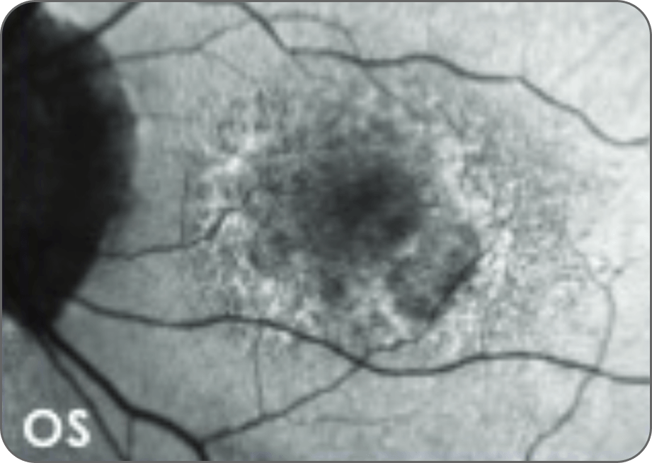
BASELINE
BCVA 20/25
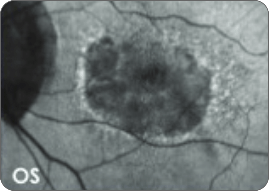
~1 YEAR
BCVA 20/40
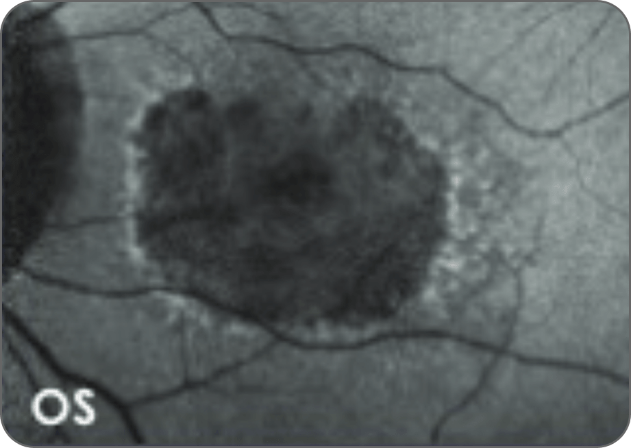
~1.5 YEARS
BCVA 20/400 (no near VA)
Real–world case of a 75–year–old female and images courtesy of Mary Beth Yackey, OD (individual experiences may vary).4
BCVA is weakly correlated to GA lesion progression, offering limited prognostic value1,5-8
Microperimetry may provide a more accurate assessment of functional vision to evaluate the impact of disease and/or treatment1,5,6,9,10
BCVA=best-corrected visual acuity; IRIS=Intelligent Research in Sight; nAMD=neovascular age-related macular degeneration; NSF=nonsubfoveal; SF=subfoveal; VA=visual acuity.
Understanding COMPLEMENT
Complement overactivation is strongly associated with GA progression11,12
Increased levels of complement activity have been found in GA lesions and surrounding areas, including photoreceptors. C3 is the central protein in the complement cascade. Three complement activation pathways converge when C3 splits into C3a and C3b.13-15
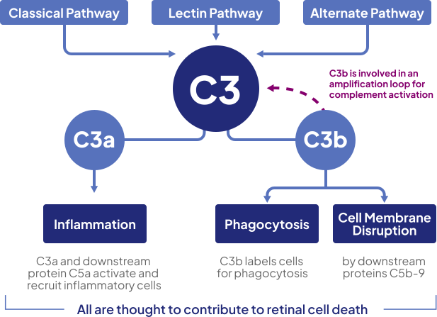
C3 overactivation unleashes negative effects downstream, resulting in retinal cell death14,15
-
Why is early intervention, such as early diagnosis and ongoing monitoring, critical in GA?
Vision lost to GA progression can never be fully recovered and can harm multiple aspects of your patients' lives.1,2
Learn more -
How rapidly does GA vision loss progress?
GA vision loss progresses faster than you may think. In fact, ~1 in 5 patients with GA become legally blind in their better–seeing eye within 1 year.3
Learn more- Based on a retrospective analysis of clinical data recorded in the IRIS® Registry of electronic health records from 69,441 patients diagnosed with GA in 1 eye and GA or nAMD in the other eye (mean baseline VA was 63 letters [bilateral GA], 56 letters [unilateral GA]) between January 1, 2016 and December 31, 2017. Patients diagnosed with nAMD <3 years prior to GA in the study eye were excluded. N-values and percentages were calculated from SF and NSF patient subset data.3
-
What are the most common early warning signs of GA?
Screening for GA often starts by knowing the key risk factors for the development of GA, including family history, history of smoking, diet, obesity, certain dyslipidemias, cardiovascular disease, and clinical findings from imaging such as GA in fellow eye or increased drusen volume. Patients with GA may experience straight lines that appear distorted, dull or washed-out colors, blurred vision, difficulty seeing in low light, reduced central vision, or missing spots in vision.1,18-21
Learn more
- Fleckenstein M, Mitchell P, Freund KB, et al. The progression of geographic atrophy secondary to age-related macular degeneration. Ophthalmology. 2018;125(3):369-390.
- Bakri SJ, Brinkmann CK, Mulvey A, et al. Characterizing patient perceptions of living with geographic atrophy: the global geographic atrophy insights survey. Clin Ophthalmol. 2024;18:3725–3737.
- Rahimy E, Ali Khan M, Ho AC, et al. Progression of geographic atrophy: retrospective analysis of patients from the IRIS® Registry (Intelligent Research in Sight). Ophthalmology Sci. 2023;3(4):100318. doi:10.1016/j.xops.2023.100318
- Data on file. Apellis Pharmaceuticals, Inc.
- Kim JE. Retinal sensitivity following pegcetacoplan treatment for geographic atrophy in the GALE study. Presented at: Macula Society Meeting 2025; February 12-15, 2025; Charlotte Harbor, FL.
- Csaky KG, Patel PJ, Sepah YJ, et al. Microperimetry for geographic atrophy secondary to age-related macular degeneration. Surv Ophthalmol. 2019;64(3):353-364.
- Anegondi N, Steffen V, Sadda SR, et al. Visual loss in geographic atrophy: learnings from the lampalizumab trials. Ophthalmology. 2025;132(4):420-430.
- Meleth AD, Mettu P, Agrón E, et al. Changes in retinal sensitivity in geographic atrophy progression as measured by microperimetry. Invest Ophthalmol Vis Sci. 2011;52(2):1119-1126.
- Heier JS, Pieramici D, Chakravarthy U, et al. Visual function decline resulting from geographic atrophy: results from the Chroma and Spectri phase 3 trials. Ophthalmol Retina. 2020;4(7):673-688.
- Chakravarthy U, Schwartz R, Guymer RH, et al. Visual function benefit after treatment with pegcetacoplan: microperimetry analysis from the phase 3 Oaks trial. Am J Ophthalmol. 2025;273:119-129.
- Holz FG, Strauss EC, Schmitz-Valckenberg S, van Lookeren Campagne M. Geographic atrophy: clinical features and potential therapeutic approaches. Ophthalmology. 2014:121(5):1079-1091.
- van Lookeren Campagne M, LeCouter J, Yaspan BL, Ye W. Mechanisms of age-related macular degeneration and therapeutic opportunities. J Pathol. 2014:232(2):757-764.
- Katschke KJ Jr, Xi H, Cox C, et al. Classical and alternative complement activation on photoreceptor outer segments drives monocyte-dependent retinal atrophy. Sci Rep. 2018;8(1):7348. doi:10.7038/s41598-018-25557-8
- Smailhodzic D, Klaver CCW, Klevering BJ, et al. Risk alleles in CFH and ARMS2 are independently associated with systemic complement activation in age-related macular degeneration. Ophthalmology. 2012;119(2):339-346.
- Yates JR, Sepp T, Matharu BK, et al. Complement C3 variant and the risk of age-related macular degeneration. N Engl J Med. 2007;357(6):553-561.
- Merle NS, Church SE, Fremeaux-Bacchi V, Roumenina LT. Complement system part I - molecular mechanisms of activation and regulation. Front lmmunol. 2015;6:262. doi:10.3389/fimmu.2015.00262
- Mastellos DC, Reis ES, Ricklin D, Smith RJ, Lambris JD. Complement C3-targeted therapy: replacing long-held assertions with evidence-based discovery. Trends Immunol. 2017;38(6):383-394.
- Sobrin L, Seddon JM. Nature and nurture- genes and environment- predict onset and progression of macular degeneration. Prog Retin Eye Res. 2024;40:1-15.
- Lad EM, Finger RP, Guymer R. Biomarkers for the progression of intermediate age-related macular degeneration. Ophthalmol Ther. 2023;12(6):2917-2941.
- Sacconi R, Corbelli E, Querques L, et al. A review of current and future management of geographic atrophy. Ophthalmol Ther. 2017;6(1):69-77.
- Schultz NM, Bhardwaj S, Barclay C, Gaspar L, Schwartz J. Global burden of dry age-related macular degeneration: a targeted literature review. J Clin Ther. 2021;43(10):1792-1818.