Monthly (n=202)

SYFOVRE Efficacy
Study Design
SYFOVRE has an unmatched amount of clinical evidence in patients with GA secondary to AMD1-4
Studied over 5 years in >1200 patients1,2,5,6
OAKS and DERBYTwo 2-year, double-masked trials
GALE 3-year, open-label extension trial
83% of patients continued to GALE*†
SYFOVRE monthly (n=419)
SYFOVRE monthly (n=241)
SYFOVRE EOM (n=420)
SYFOVRE EOM (n=238)
Sham monthly (n=208)
SYFOVRE monthly (n=129)
Sham EOM (n=211)
SYFOVRE EOM (n=144)
- Patients received 15 mg/0.1 mL intravitreal SYFOVRE monthly or EOM.1
- First GALE visit was required to be within 60 days of the final OAKS and DERBY visit.6
- Ten patients from Phase 1b Study 103 enrolled in GALE and are included in the safety analysis.6
SYFOVRE has the largest and longest clinical data set of any approved GA treatment1,6-9
SYFOVRE is the only approved GA treatment with efficacy and safety evaluated for 5 years1-3,6
Up to 2 years1:
OAKS and DERBY
Primary assessment: changes from baseline in rate of GA lesion area growth measured by FAF
Up to 5 years2:
GALE
Primary and secondary assessments: incidence and severity of ocular and systemic AEs, and change from baseline in total area of GA lesion(s) in the study eye (mm2)
GALE trial limitations2,6:
GALE was a long-term, multicenter, Phase 3 open-label extension study, subject to patient dropouts over time. The analysis used a projected sham (based on Years 0–2 sham data) which may not reflect the rate of change for all GA patients. Although analyses were prespecified, there was no statistical testing hierarchy, so results should be interpreted with caution. Open-label design may introduce selection bias. Efficacy data presented for GALE are in patients with NSF lesions at baseline in OAKS and DERBY.
Discontinuation rate through 2 years (SYFOVRE monthly vs SYFOVRE EOM vs sham pooled, respectively)1:
- 31% vs 21% vs 25% in OAKS
- 29% vs 22% vs 21% in DERBY
Discontinuation rate over 5 years (SYFOVRE monthly vs SYFOVRE EOM vs pooled crossover from sham)6:
- 29% vs 33% vs 37% in GALE
AE=adverse event; AMD=age-related macular degeneration; EOM=every other month; FAF=fundus autofluorescence; GA=geographic atrophy; NSF=nonsubfoveal.
Baseline patients and study eye characteristics6
OAKS |
DERBY |
|||||
|---|---|---|---|---|---|---|
| SYFOVRE monthly (n=202) |
SYFOVRE EOM (n=205) |
Sham pooled (n=207) |
SYFOVRE monthly (n=201) |
SYFOVRE EOM (n=201) |
Sham pooled (n=195) |
|
| PATIENT CHARACTERISTICS | ||||||
| Mean age (years) | 79 | 78 | 79 | 79 | 79 | 79 |
| Female | 62% | 57% | 64% | 59% | 60% | 63% |
| Male | 38% | 43% | 36% | 41% | 40% | 37% |
| Caucasian | 92% | 92% | 91% | 93% | 93% | 96% |
| Fellow eye CNV | 21% | 18% | 21% | 19% | 20% | 19% |
| GEOGRAPHIC REGION | ||||||
| US | 73% | 69% | 71% | 71% | 61% | 63% |
| ROW | 27% | 31% | 29% | 29% | 39% | 37% |
| STUDY EYE CHARACTERISTICS | ||||||
| Mean GA lesion size | 8.2 mm² | 8.3 mm² | 8.2 mm² | 8.4 mm² | 8.3 mm² | 8.2 mm² |
| GA lesion size <7.5 mm² | 50% | 48% | 50% | 49% | 49% | 49% |
| GA lesion location without subfoveal involvement | 43% | 36% | 29% | 36% | 40% | 37% |
| GA lesion focality unifocal | 29% | 30% | 33% | 27% | 26% | 34% |
| Bilateral GA | 83% | 85% | 80% | 82% | 80% | 77% |
| Number of intermediate/large drusen >20 | 46% | 51% | 50% | 39% | 39% | 50% |
| Pseudodrusen (NIR) | 83% | 87% | 84% | 89% | 90% | 85% |
| Mean NL-BCVA score | 61 | 58 | 58 | 60 | 59 | 59 |
| Mean LLD*—ETDRS letters | 27 | 26 | 25 | 27 | 26 | 26 |
OAKS |
|||
|---|---|---|---|
| SYFOVRE monthly (n=202) |
SYFOVRE EOM (n=205) |
Sham pooled (n=207) |
|
| PATIENT CHARACTERISTICS | |||
| Mean age (years) | 79 | 78 | 79 |
| Female | 62% | 57% | 64% |
| Male | 38% | 43% | 36% |
| Caucasian | 92% | 92% | 91% |
| Fellow eye CNV | 21% | 18% | 21% |
| GEOGRAPHIC REGION | |||
| US | 73% | 69% | 71% |
| ROW | 27% | 31% | 29% |
| STUDY EYE CHARACTERISTICS | |||
| Mean GA lesion size | 8.2 mm² | 8.3 mm² | 8.2 mm² |
| GA lesion size <7.5 mm² | 50% | 48% | 50% |
| GA lesion location without subfoveal involvement | 43% | 36% | 29% |
| GA lesion focality unifocal | 29% | 30% | 33% |
| Bilateral GA | 83% | 85% | 80% |
| Number of intermediate/large drusen >20 | 46% | 51% | 50% |
| Pseudodrusen (NIR) | 83% | 87% | 84% |
| Mean NL-BCVA score | 61 | 58 | 58 |
| Mean LLD*—ETDRS letters | 27 | 26 | 25 |
DERBY |
|||
|---|---|---|---|
| SYFOVRE monthly (n=201) |
SYFOVRE EOM (n=201) |
Sham pooled (n=195) |
|
| PATIENT CHARACTERISTICS | |||
| Mean age (years) | 79 | 79 | 79 |
| Female | 59% | 60% | 63% |
| Male | 41% | 40% | 37% |
| Caucasian | 93% | 93% | 96% |
| Fellow eye CNV | 19% | 20% | 19% |
| GEOGRAPHIC REGION | |||
| US | 71% | 61% | 63% |
| ROW | 29% | 39% | 37% |
| STUDY EYE CHARACTERISTICS | |||
| Mean GA lesion size | 8.4 mm² | 8.3 mm² | 8.2 mm² |
| GA lesion size <7.5 mm² | 49% | 49% | 49% |
| GA lesion location without subfoveal involvement | 36% | 40% | 37% |
| GA lesion focality unifocal | 27% | 26% | 34% |
| Bilateral GA | 82% | 80% | 77% |
| Number of intermediate/large drusen >20 | 39% | 39% | 50% |
| Pseudodrusen (NIR) | 89% | 90% | 85% |
| Mean NL-BCVA score | 60 | 59 | 59 |
| Mean LLD*—ETDRS letters | 27 | 26 | 26 |
LLD at baseline was available for 199, 199, and 192 patients in the monthly, EOM, and sham groups, respectively, in DERBY.6
A broad range of patients were included6
Key inclusion criteria5,6:
- GA lesion requirements
- Total size: 2.5 mm2 to 17.5 mm2
- With and without SF lesion involvement
- If multifocal, at least 1 focal lesion must be ≥1.25 mm2
- Presence of perilesional hyperautofluorescence
- Age ≥60 years
- NL-BCVA ≥24 letters using ETDRS charts in the study eye
Key exclusion criteria6:
- GA secondary to a condition other than AMD such as Stargardt disease in either eye
- History of or active CNV in the study eye, including presence of RPE rips (assessed by reading center)
- Prior participation in any other interventional clinical study for GA in either eye
CNV=choroidal neovascularization; ETDRS=Early Treatment Diabetic Retinopathy Study;
LLD=low-luminance deficit; NIR=near-infrared reflectance; NL-BCVA=normal luminance best-corrected
visual acuity; ROW=rest of world; RPE=retinal pigment epithelium.
Among agents approved to treat GA, only SYFOVRE was studied in trials that allowed patients with SF
lesions in the study eye or CNV in the fellow eye4-6,10
Explore the latest results from the GALE extension trial below
ANATOMICAL DATA
Based on clinical trials in Patients with GA
Only SYFOVRE demonstrated results in SF and NSF lesions1,2,4,5,10
Reduced the rate of GA lesion area growth with monthly or EOM dosing vs sham pooled (measured by FAF at Year 2)1
OAKS1
-
-
EOM (n=205)
18%
-0.72 mm2 (95% Cl: -1.10 to -0.33)
DERBY1
-
Monthly (n=201)
18%
-0.73 mm2 (95% Cl: -1.14 to -0.31) -
EOM (n=201)
17%
-0.70 mm2 (95% Cl: -1.11 to -0.28)
SYFOVRE has evidence in patients commonly seen in clinical practice1,2,4,5,10
-
GA patients with SF or NSF lesions in the study eye or CNV in the fellow eye were enrolled in all Phase 3 clinical trials1,2,4,10
- SF lesions typically grow at a slower rate than NSF lesions2
- Patients with GA secondary to a condition other than AMD and history or presence of CNV in the study eye were excluded5
SF=subfoveal.
In an analysis of the first 2 years vs the last 3 years of treatment (NSF PATIENTS)
SYFOVRE effects strengthened over time6
Increasing reductions in lesion growth rate (Years 0-2 vs 2-5)
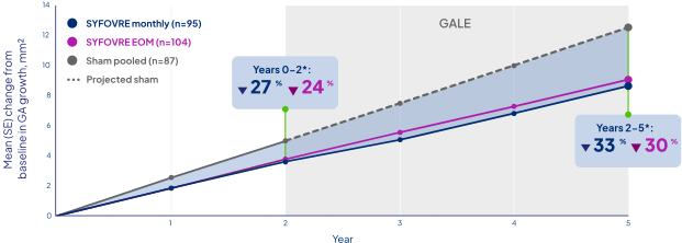
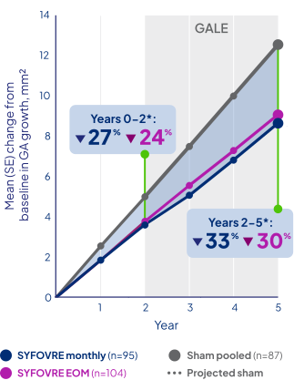
Projected sham from Year 2 to Year 5 was estimated from the mean rate of change in each 6-month period from Year 0 to Year 2 in the sham group.2,11
Overall reduction in Years 0-5 was 27% (-3.4 mm2) and 31% (-3.9 mm2) for EOM and monthly doses, respectively, in NSF patients.6
These are prespecified analyses with no statistical testing planned, and no conclusions can be drawn.6
SYFOVRE is the only treatment with 5 years of clinical
trial evidence in GA1–3,6
These are prespecified analyses with no statistical testing planned, and no conclusions can be drawn.6
FDA=US Food and Drug Administration. In a PRESPECIFIED analysis of GALE that assumed a linear growth rate for projected sham (NSF PATIENTS)2,6
Starting SYFOVRE sooner showed more retinal tissue was saved6,11
More tissue preservation observed with earlier treatment (equivalent to ~1.5 optic discs)
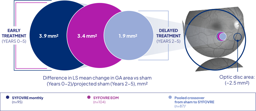
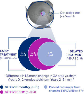
Pooled crossover included patients from both monthly and EOM sham arms in OAKS and DERBY who crossed over into SYFOVRE monthly and EOM treatment in GALE, respectively.2,6
This is a prespecified analysis with no statistical testing planned, and no conclusions can be drawn.6
LS=least squares. Examining ellipsoid zone data provides a precise view of photoreceptor loss12
OCT-based AI analysis revealed a greater loss of ellipsoid zone layer than RPE12
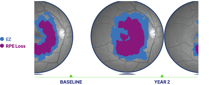
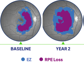
For illustrative purposes only. OCT-based evaluation of GA lesion progression in an untreated eye. Adapted from Schmidt-Erfurth U, et al; Ophthalmology; 2025; with permission from Elsevier.
- The photoreceptor layer is represented on an OCT scan by the EZ layer12
- EZ layer loss is significantly correlated to GA lesion growth in observational studies12,13
Ellipsoid zone loss precedes the loss of RPE12
AI=artificial intelligence; EZ=ellipsoid zone; OCT=optical coherence tomography.
In a post hoc analysis of patients in the OAKS and DERBY trials12
Photoreceptor preservation was observed in SYFOVRE patients12
Reduction in the rate of ellipsoid zone layer loss was observed with SYFOVRE vs sham pooled at Year 2
OAKS12
-
Monthly
53%
-
EOM
46%
DERBY12
-
Monthly
47%
-
EOM
46%
An analysis of 897 eyes (mean baseline lesion 8.2 mm2 by FAF) from 897 patients who were enrolled in the OAKS and DERBY trials, imaged with a Spectralis OCT, evaluating the change in EZ layer area.12
Limitations:
- This is a post hoc analysis with no control for type 1 error and should be interpreted with caution6
- Analysis only included subjects examined with Spectralis OCT12
- Analysis does not include subjects whose GA lesions expanded beyond OCT field of view12
- AI algorithm has not been approved by the FDA
Methodology:
- Volumetric Spectralis OCT scans were analyzed using a deep learning algorithm that segmented the EZ and inner RPE boundaries to calculate EZ–RPE thickness. Thickness values were mapped across the macula to quantify localized photoreceptor loss. EZ loss was defined as an EZ–RPE thickness of ≤4 μm12
Ellipsoid zone data offer insight into photoreceptor preservation12
Functional DATA
Microperimetry is a comprehensive assessment of functional vision in GA14-17
A visual field test of macular sensitivity14–17
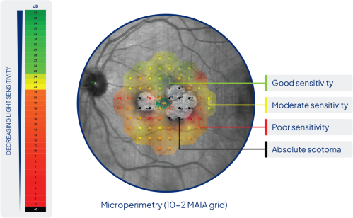
Microperimetry color maps are interpolated based on sensitivity at the 68 microperimetry points. They may not represent the sensitivity of all areas in the color map.
How it works:
- Assesses patient-reported perception to light at variable intensity (-1 dB to 36 dB)14,15,17
- Analyzes the extent of scotoma at different areas around the fovea15,16
Why it works:
- Highest correlation with GA area compared with BCVA, LLVA, NEI VFQ-25, FRI index, and reading speed14,18
Limitation:
Patient reliability and fixation are potential limitations of microperimetry.
Microperimetry has the strongest correlation to retinal structure and function of GA clinical trial endpoints14,18
BCVA=best-corrected visual acuity; dB=decibels; FRI=functional reading independence; LLVA=low-luminance visual acuity; MAIA=macular integrity assessment; NEI VFQ=National Eye Institute Visual Function Questionnaire.
In a post hoc microperimetry analysis vs sham pooled through 2 years (OAKS trial)
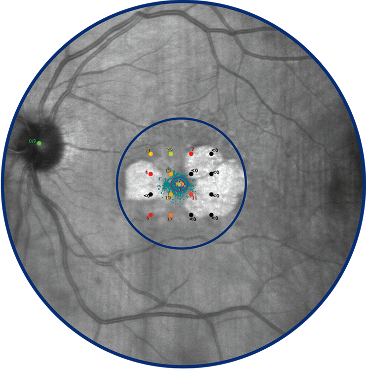
Reduced risk of scotomas in the central macular area was observed with SYFOVRE6,14
Absolute scotoma in central 16 loci was assessed by microperimetry at Year 2 (n=614)6
-
Monthly reduced risk6,14
43%
(HR=0.57;95% Cl: 0.33 to 0.98) -
EOM reduced risk6,14
48%
(HR=0.52;95% Cl: 0.32 to 0.85)
Location matters. The central macular area determines central vision, which significantly impacts daily living (eg, recognizing faces, reading, driving).15,17,19
Important contextual information:
- This analysis has no control for type 1 error, and no conclusions can be drawn6
- In the OAKS and DERBY trials, there was no statistically significant difference in prespecified key secondary endpoints on functional measures (eg, BCVA, reading speed, FRI index, microperimetry mean sensitivity [OAKS only]) at 2 years6
- Functional outcomes declined similarly in all 3 treatment groups over 2 years, consistent with the natural history of the disease6
-
How many SYFOVRE® (pegcetacoplan) clinical trials have been conducted?
SYFOVRE has an unmatched amount of clinical evidence in GA, with the largest and longest clinical data set of any GA treatment. The clinical trials of SYFOVRE include two 2–year double-masked trials (OAKS and DERBY) and the 3–year open-label GALE extension trial.1-4
Learn more -
How many patients were included in the SYFOVRE (pegcetacoplan injection) clinical trials?
SYFOVRE was studied in clinical trials over 5 years in >1200 patients with GA secondary to AMD with either SF or NSF lesions.1,2
Learn more -
What treatment effects were assessed in the 5-year GALE open-label extension study?
GALE measured retinal tissue preservation and slowing of GA lesion growth over 5 years vs projected sham.2,6
Learn more -
In which patient population was SYFOVRE (pegcetacoplan injection) studied?
Patients with GA secondary to AMD with SF or NSF lesions in the study eye or CNV in the fellow eye were enrolled in all Phase 3 clinical trials1,2,4,10
- SF lesions typically grow at a slower rate than NSF lesions
- Patients with GA secondary to a condition other than AMD and history or presence of CNV in the study eye were excluded
-
Does SYFOVRE (pegcetacoplan injection) have tissue preservation data?
Yes, tissue preservation was evaluated over 5 years in GALE.6,11
Learn more -
What discontinuation rates were seen in the clinical trials of SYFOVRE (pegcetacoplan injection)?
In clinical trials of SYFOVRE, discontinuation rates through 2 years (SYFOVRE monthly vs SYFOVRE every other month vs sham pooled, respectively) were 31% vs 21% vs 25% in OAKS and 29% vs 22% vs 21% in DERBY.1
Discontinuation rates (SYFOVRE monthly vs SYFOVRE EOM vs pooled crossover from sham) were 29% vs 33% vs 37% over 5 years in GALE.6
Learn more
- Syfovre. Package insert. Apellis Pharmaceuticals, Inc.; 2025.
- Wykoff CC, Holz FG, Chiang A, et al; OAKS, DERBY, and GALE investigators. Pegcetacoplan treatment for geographic atrophy in age-related macular degeneration over 36 months: data from OAKS, DERBY, and GALE. Am J Ophthalmol. 2025;276:350-364.
- Izervay. Package insert. Astellas Pharma US, Inc.; 2025.
- Patel SS, Lally DR, Hsu J, et al. Avacincaptad pegol for geographic atrophy secondary to age-related macular degeneration: 18-month findings from the GATHER1 trial. Eye (Lond). 2023;37(17):3551-3557.
- Heier JS, Lad EM, Holz FG, et al; OAKS and DERBY study investigators. Pegcetacoplan for the treatment of geographic atrophy secondary to age-related macular degeneration (OAKS and DERBY): two multicentre, randomised, double-masked, sham-controlled, phase 3 trials. Lancet. 2023;402(10411):1434-1448.
- Data on file. Apellis Pharmaceuticals, Inc.
- Jaffe GJ, Westby K, Csaky KG, et al. C5 inhibitor avacincaptad pegol for geographic atrophy due to age-related macular degeneration: a randomized pivotal phase 2/3 trial. Ophthalmology. 2021;128(4):576-586.
- A phase 3 safety and efficacy study of intravitreal administration of Zimura (complement C5 inhibitor). ClinicalTrials.gov identifier NCT04435366. Updated October 21, 2025. Accessed February 5, 2026. https://clinicaltrials.gov/study/NCT04435366
- Holz FG, Sadda SR, Busbee B, et al. Efficacy and safety of lampalizumab for geographic atrophy due to age-related macular degeneration: Chroma and Spectri phase 3 randomized clinical trials. JAMA Ophthalmol. 2018;136(6):666-677.
- Khanani AM, Patel SS, Staurenghi G, et al; GATHER2 trial investigators. Efficacy and safety of avacincaptad pegol in patients with geographic atrophy (GATHER2): 12-month results from a randomised, double-masked, phase 3 trial. Lancet. 2023;402(10411):1449-1458.
- Singerman LJ; OAKS, DERBY, and GALE investigators. 24-month results from the GALE open-label extension study: 48 months of continuous pegcetacoplan. Presented at: Macula Society Meeting 2025; February 12-15, 2025; Charlotte Harbor, FL.
- Schmidt-Erfurth U, Mai J, Reiter GS, et al. Disease activity and therapeutic response to pegcetacoplan for geographic atrophy identified by deep learning-based analysis of OCT. Ophthalmology. 2025;132(2):181-193.
- Coulibaly LM, Reiter GS, Fuchs P, et al. Progression dynamics of early versus later stage atrophic lesions in nonneovascular age-related macular degeneration using quantitative OCT biomarker segmentation. Ophthalmol Retina. 2023;7(9):762-770.
- Kim JE. Retinal sensitivity following pegcetacoplan treatment for geographic atrophy in the GALE study. Presented at: Macula Society Meeting 2025; February 12-15, 2025; Charlotte Harbor, FL.
- Csaky KG, Patel PJ, Sepah YJ, et al. Microperimetry for geographic atrophy secondary to age-related macular degeneration. Surv Ophthalmol. 2019;64(3):353-364.
- Meleth AD, Mettu P, Agrón E, et al. Changes in retinal sensitivity in geographic atrophy progression as measured by microperimetry. Invest Ophthalmol Vis Sci. 2011;52(2):1119-1126.
- Chakravarthy U, Schwartz R, Guymer RH, et al. Visual function benefit after treatment with pegcetacoplan: microperimetry analysis from the phase 3 Oaks trial. Am J Ophthalmol. 2025;273:119-129.
- Heier JS, Pieramici D, Chakravarthy U, et al. Visual function decline resulting from geographic atrophy: results from the Chroma and Spectri phase 3 trials. Ophthalmol Retina. 2020;4(7):673-688.
- Koksaldi S, Kayabasi M, Karti O, Saatci AO. Clinical anatomy of the macula. Med Hypothesis Discov Innov Ophthalmol. 2025;14(2):17-27.